
10 Tips for Choosing the Best 3D Skin Scanner
A 3D Skin Scanner can turn a quick consultation into a detailed visual record. It maps contours, captures surface features, and helps compare images over time. But polished graphics alone do not guarantee useful results. Lighting, patient positioning, software, and operator training all affect what the scanner shows.
Dermatologist and aesthetic-medicine researcher Dr. Michael H. Gold is a recognized voice in clinical skin care. A practical principle for evaluating scanners is: “Consistent images matter more than impressive graphics.” Treat this as a buying criterion, not a substitute for clinical judgment. Ask vendors how they control lighting and positioning, whether scans can be repeated reliably, and how the system stores and compares results. See the details.
This guide’s ten tips focus on those real-world checks. Look closely at image clarity, measurement repeatability, workflow, training, support, and total ownership costs. Request a live demonstration. Watch how long a scan takes, how the patient is positioned, and whether the results are easy to explain. A scanner should fit your clinic’s needs, not just its showroom. Consider what it measures, what it cannot measure, and how staff will communicate its findings. That distinction matters.
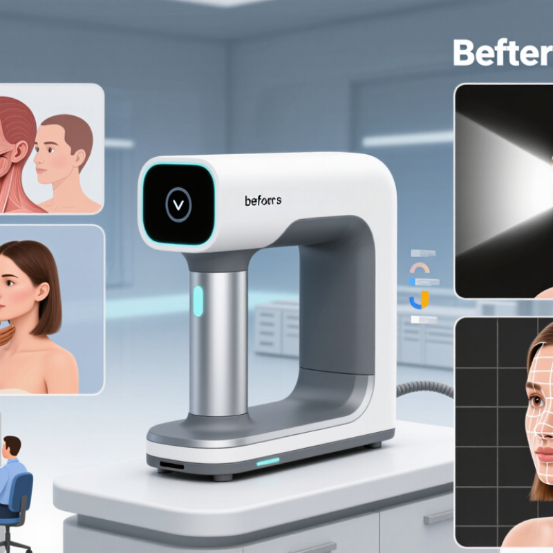
Understanding the Purpose and Applications of a 3D Skin Scanner
A 3D skin scanner captures a measurable surface record, not merely a flattering photograph. Clinics may use it to document wrinkles, pores, pigmentation, redness, and facial contours before and after treatment. The World Health Organization reported in 2023 that skin conditions affect about 1.8 billion people at any given time. That scale makes consistent assessment valuable, but a scanner cannot diagnose disease or replace a clinician’s examination. Not a diagnosis. Results can also shift with room lighting, camera distance, skin preparation, and device calibration. A precise-looking image is not automatically a precise measurement.
Tips: Match the scanner to its intended use. For treatment follow-up, check whether it saves repeatable images and tracks the same regions over time. Ask how it performs across different skin tones, and request evidence on measurement repeatability—not just sample images. Keep the room lighting and patient positioning consistent; a chin tilted slightly upward can change contour readings. Record product use or recent procedures too. These details are easy to overlook. They can explain an apparent change that the scan alone cannot. A clinician should interpret the images alongside symptoms, medical history, and direct examination.
10 Tips for Choosing the Best 3D Skin Scanner - Understanding the Purpose and Applications of a 3D Skin Scanner
| No. | Selection Tip | What to Evaluate | Relevant Applications | Questions to Ask |
|---|---|---|---|---|
| 1 | Define the intended purpose | Clarify whether the system is intended for clinical assessment, aesthetic consultation, research, treatment follow-up, or another defined workflow. | Dermatology, aesthetic practices, skin-care consultations, and research settings. | Which specific decisions or tasks should the scan support? |
| 2 | Check the measurements and outputs | Review what the system captures, such as surface shape, colour images, texture-related features, or mapped areas. Capabilities differ between systems. | Documenting visible skin features, comparing scans, and supporting consultations. | Which outputs are directly measured, and which are estimates or software-generated indicators? |
| 3 | Assess scan consistency | Look for clear guidance on positioning, lighting, distance, facial expression, and repeat-scan procedures. Consistent conditions help make comparisons more meaningful. | Before-and-after documentation and longitudinal monitoring. | How does the system help users reproduce the same capture conditions? |
| 4 | Evaluate coverage and capture time | Confirm which areas can be scanned, whether multiple views are needed, and how long capture and processing typically take in your workflow. | Full-face assessment, targeted-area documentation, and patient consultations. | Can the intended areas be captured comfortably and efficiently? |
| 5 | Review image quality and detail | Inspect sample scans for focus, consistent colour, visible surface detail, and areas that may be obscured by hair, reflections, or movement. | Visual documentation, case review, and research image collection. | Can you review representative scans from different skin tones and skin conditions? |
| 6 | Understand validation and limitations | Ask for evidence supporting the system’s stated measurements and intended uses. A visualisation or software score should not automatically be treated as a diagnosis. | Clinical support, research, and non-diagnostic aesthetic assessment. | What has been independently evaluated, and what limitations should users understand? |
| 7 | Check workflow and software fit | Review how scans are stored, compared, exported, and incorporated into existing procedures. Confirm compatibility requirements before purchase. | Practice documentation, team review, and research data management. | Does the software support the file formats and workflow your team needs? |
| 8 | Review privacy and data security | Determine how identifiable images and records are collected, accessed, stored, backed up, and deleted. Follow applicable privacy rules in your location. | Any setting that captures identifiable patient or participant images. | Who can access the data, where is it stored, and how is consent documented? |
| 9 | Consider accessibility and ease of use | Assess setup, operator training, patient comfort, accessibility needs, and the clarity of instructions and reports. | Busy clinics, multi-user teams, and repeated follow-up visits. | Can staff learn the process, and can patients complete it comfortably? |
| 10 | Compare total ownership costs and support | Include the scanner, software, maintenance, training, updates, data storage, and any required accessories when comparing costs. | Long-term use in clinical, commercial, or research environments. | What ongoing costs, service arrangements, and technical support are included? |
Note: 3D skin scanner functions and measurement methods vary. Confirm that a system is suitable for its intended use, review its evidence and limitations, and follow applicable clinical, privacy, and regulatory requirements.

© 2025 EXCELSIUS MEDICAL All rights reserved